


Figure. Ventral and dorsal views of Mp. hjorti, 48 mm ML, captured from the Johnson Sea Link II submersible at 24°02'N, 81°54'W at a depth of 910 m, preserved. Photographs by R. Young
Characteristics
- Arms
- Large arm suckers with 9- 10 truncated teeth on distal margin (Chun, 1913).
- Large arm suckers 1.3 mm in diameter from 95 mm ML specimen (Chun, 1913).
- Tentacles
- Club not thickened compared to stalk; trabeculate protective membranes well developed (Chun, 1913).
- Club suckers 0.25 mm in diameter from squid 68 mm ML (Chun, 1913).
- Club suckers arranged in two irregular rows proximally gradually increasing to 18-20 in an oblique row (Chun, 1913).
- Head
- Deep pocket between funnel bridles absent.
- Mantle and Skin
- Large tubercules cover mantle, head, funnel and aboral surface of arms in subadults.
- Pigmentation
- Color a deep red. Pigment mostly in chromatophores with some underlying dermal pigmentation.
- Measurements (in mm) and counts
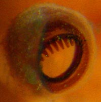
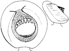
Figure. Arm suckers of Mp. hjorti. Left - Distal-oblique view, 48 mm ML, 24°02'N, 81°54'W. Photograph by R. Young. Right - Oral and side views, lateral arm, Gulf of Guinea. Drawing from Rancurel (1973).
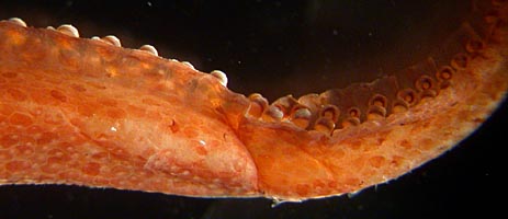
Figure. Lateral view of an arm of Mp. hjorti, showing protective membranes, 48 mm ML, preserved.
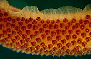

Figure. Left - Oral view of a portion of the tentacular club of Mp. hjorti, 48 mm ML, preserved. Photograph by R. Young. Right - Oral view of a club sucker of Mp. hjorti, 10°04'N, 18°22'W, tentacle collected without squid. Photographs by R. Young.
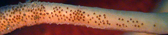
Figure. Oral view of the base of the tentacular club of Mp. hjorti, 48 mm ML, preserved. Photograph by R. Young.
Scanning electron micrographs of the suckers of Mp. hjorti can be seen here.
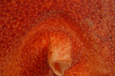

Figure. Left - Ventral view of the funnel and posterior region of the head of Mp. hjorti showing the absence of a pocket between funnel bridles, 48 mm ML. Right - Ventral view of the olfactory organ of Mp. hjorti, same squid. Photographs by R. Young.
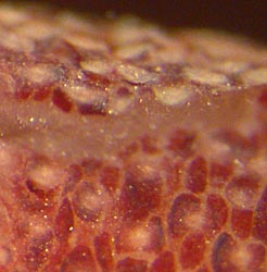
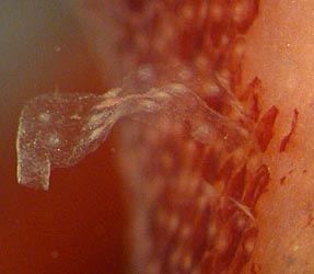
Figure. Tubercules of Mp. hjorti, 48 mm ML. Left - Ventral view of a small region at the side of the head showing tubercules from two different angles. Right - Region near an arm base showing an apparent detached cuticle with tubercules that formerly covered the skin. Compare with photographs higher up on this page and on the species page. Photographs by R. Young.
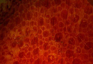
Figure. Ventral view of a portion of the fin (?) of Mp. hjorti showing chromatophores. 48 mm ML, preserved. Photograph by R. Young.
| Source | Chun, 1913 | Rancurel, 1973 | Rancurel, 1973 |
| Mantle length | 95 | 44 | 40 |
| Mantle width | 35 | 11 | 12 |
| Fin length | -- | 39 | 36 |
| Fin width | 102 | 51 | 51 |
| Head width | 41 | -- | -- |
| Eye diameter | 26 | 10 | 9 |
| Arm I, length | 44 | 17 | 13 |
| Arm II, length | -- | -- | 19 |
| Arm III, length | -- | -- | 14 |
| Arm IV, length | 74 | -- | -- |
| Arm I, sucker pairs | -- | -- | 27 |
| Arm II, sucker pairs | -- | -- | 40 |
| Arm III, sucker pairs | -- | -- | 29 |
| Club length | 75 | -- | -- |


